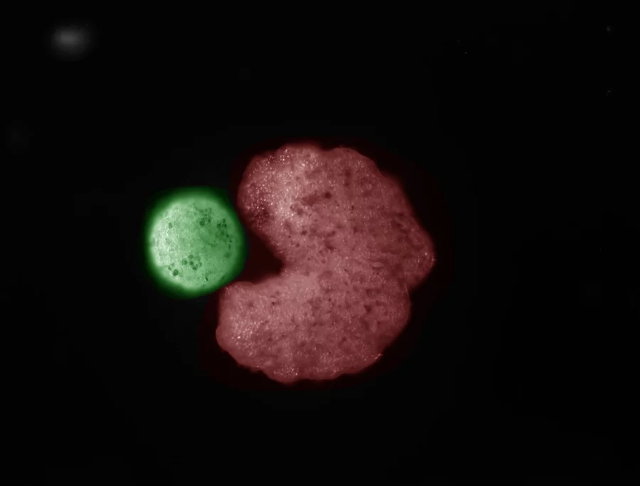
Scientists Develop Solution that Renders Living Skin Transparent
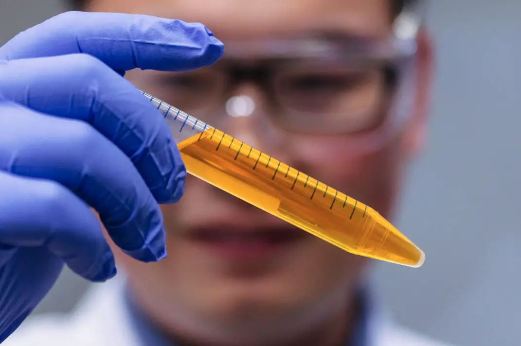
- Research suggests scientists have made living mouse skin transparent using tartrazine, a common food dye.
- The process is reversible, safe, and could revolutionize biomedical imaging.
- It seems likely that human applications are possible, but challenges remain due to thicker skin.
- An unexpected detail is that this technique allows real-time observation of internal organs without invasive procedures.
In a pioneering study published on September 5, 2024, in the journal Science, researchers led by Dr. Zihao Ou, now an assistant professor of physics at The University of Texas at Dallas (UT Dallas), have developed a revolutionary technique to make the skin of living mice transparent using tartrazine, a common yellow food coloring also known as FD&C Yellow #5.
This discovery, detailed in various reports from UT Dallas, Stanford, and other reputable sources, marks a significant advancement in biomedical imaging and research, with potential implications for human medicine.
Research Methodology and Findings
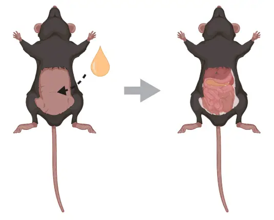
The study focused on live mice, applying a mixture of water and tartrazine to the skin on their skulls and abdomens.
The process, which takes a few minutes to achieve transparency, is reversible by washing off any remaining dye, with the diffused dye being metabolized and excreted through urine.
This ensures the technique is safe and biocompatible, as tartrazine is certified by the Food and Drug Administration (FDA) for use in foods like orange- or yellow-colored snack chips and candy coatings.
The mechanism behind this transparency is rooted in optics.
Living skin is a scattering medium, akin to fog, which scatters light due to the mismatch in refractive indices between its components, such as lipids and water.
By dissolving tartrazine in water, researchers altered the solution’s refractive index to match that of the skin’s tissue components, reducing light scattering.
This effect, described by Dr. Ou as looking “like a magic trick” to the uninitiated, is based on fundamental physics, specifically the Kramers-Kronig relations, which describe the relationship between light absorption and refractive index.
Through the transparent skin, researchers observed blood vessels on the brain’s surface, internal organs like the liver and intestines, and peristalsis—the muscle contractions moving contents through the digestive tract.
Additional experiments, as reported by Stanford, involved applying the dye to the scalp to visualize cerebral blood vessels and to hindlimbs for high-resolution imaging of muscle fibers, demonstrating versatility.
The skin took on an orangish hue during transparency, a visual indicator of the dye’s effect.
| Aspect | Details |
|---|---|
| Research Method | Applied tartrazine solution to match refractive indices, reducing light scattering, reversible by rinsing with water, effective within minutes. |
| Observations | Blood vessels, brain activity, internal organs (liver, intestines), peristalsis, muscle fibers visible without special equipment. |
| Safety | Biocompatible, FDA-certified, metabolized and excreted, no long-term effects observed in mice. |
| Publication | Science, Sept. 5, 2024, DOI: 10.1126/science.adm6869, Achieving Optical Transparency. |
Implications for Biomedical Research and Medicine
This technique has the potential to revolutionize optical imaging in biomedical research.
Currently, microscopes and other optical equipment are limited by the opacity of living tissue, but this method allows real-time observation of biological processes without invasive procedures.
For instance, researchers could watch neuronal firing in the brain or map gastrointestinal activity, as seen in experiments with fluorescent dyes.
The Stanford Report highlights potential future applications in humans, including non-invasive melanoma testing, replacing some X-rays or CT scans, easier vein finding for blood draws, and improved laser tattoo removal.
The Guardian and other sources suggest additional medical uses, such as locating injuries, monitoring digestive disorders, and diagnosing deep-seated tumors non-invasively, potentially reducing the need for invasive biopsies.
When combined with modern imaging techniques, this could enable imaging of entire mouse brains or tumors beneath centimeter-thick tissues, expanding research beyond naturally transparent animals like zebrafish.
However, challenges remain for human applications.
Human skin is approximately 10 times thicker than a mouse’s, and it’s unclear what dosage or delivery method would be necessary to penetrate the entire thickness.
Dr. Ou noted, “In human medicine, we currently have ultrasound to look deeper inside the living body,” suggesting this technique could complement existing methods but requires further development.
Future Directions and Ongoing Research
Dr. Ou, who conducted this research while a postdoctoral researcher at Stanford before joining UT Dallas in August 2024, is continuing his work at the Dynamic Bio-imaging Lab at UT Dallas.
Future steps include understanding the optimal dosage for human tissue and experimenting with other molecules, including engineered materials, that could perform more efficiently than tartrazine.
The team, including co-authors like Dr. Guosong Hong and Dr. Mark Brongersma from Stanford, aims to improve existing research methods in optical imaging, potentially making biomedical platforms more accessible and less expensive than current technologies like MRI.
The study’s funding came from federal agencies, including the National Institutes of Health, National Science Foundation, and Air Force Office of Scientific Research, as well as the Wu Tsai Neurosciences Institute at Stanford.
The researchers have applied for a patent on the technology, indicating its commercial potential.
Public Engagement and Home Experiments
For those interested in exploring this phenomenon, the National Science Foundation has created an exercise for adults to try the Yellow #5 experiment at home using raw chicken.
This hands-on activity, detailed at NSF Activity, allows enthusiasts to witness the transparency effect, fostering a deeper understanding of the underlying physics.
Watch: Revolutionary Transparent Skin Discovery
Background on Dr. Zihao Ou
Dr. Zihao Ou earned a Bachelor of Science in physics from the University of Science and Technology of China and a doctorate in materials science and engineering from the University of Illinois Urbana-Champaign.
His doctoral research focused on electron microscopy, using electron beams for high-resolution imaging of biological and nonbiological specimens.
Seeking to impact more people, he transitioned to biological imaging techniques during his postdoctoral work at Stanford, bringing his physics and materials science background to biomedical science.
His move to UT Dallas was motivated by the university’s interdisciplinary approach, where researchers in mathematics, chemistry, biology, and physics collaborate to improve biomedical applications.
This novel approach to making living skin transparent represents a significant leap forward in biomedical imaging.
By leveraging a simple, inexpensive, and safe food dye, researchers have unlocked a new window into the body, promising to enhance our understanding of biological processes and improve medical diagnostics.
While challenges remain for human applications, the potential to revolutionize research and medicine is clear, with ongoing efforts to refine and expand this technique.


