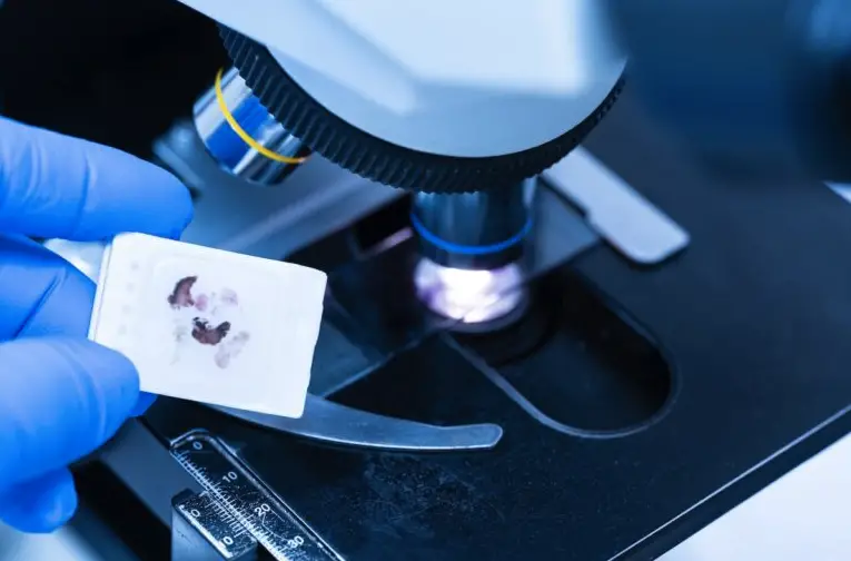
The Importance of FFPE Tissue in Histopathology: A Comprehensive Guide

Have you ever wondered how pathologists diagnose diseases from samples? Formalin-Fixed, Paraffin-Embedded (FFPE) tissue plays a pivotal role in histopathology, allowing for the detailed study of tissue architecture and cellular morphology. This technique, a cornerstone of pathology for over a century, ensures the preservation of samples for long-term analysis.
Let us explore the significance of FFPE tissue in histopathology, its preparation process, its advantages, and its applications. Understanding these aspects will highlight why this method is indispensable in diagnostic and research settings.
What Is FFPE Tissue?
It refers to samples that have been preserved using a formalin fixation process followed by embedding in paraffin wax. This method is designed to maintain the structural integrity and cellular details, which are crucial for accurate histopathological analysis.
Formalin Fixation: The first step involves immersing the sample in formalin, a formaldehyde solution in water. This process cross-links proteins, preserving the architecture and preventing degradation.
Paraffin Embedding: After fixation, the sample is dehydrated through a series of alcohol baths and then embedded in paraffin wax. This process solidifies the sample, making it easier to slice into thin sections for microscopic examination.
What Are the Advantages?
This approach offers several advantages that make it a preferred method in histopathology.
Long-term Preservation: It can be stored for decades without significant degradation, allowing for long-term studies and retrospective analyses.
Detailed Morphology: The fixation process preserves cellular details and architecture, which are essential for accurate diagnosis.
Versatility: These samples are compatible with various staining techniques, including immunohistochemistry and special stains, facilitating diverse diagnostic applications.
Cost-effectiveness: The materials and equipment required for this preparation are relatively inexpensive, making it an economical option for routine histopathological analysis.
Applications in Histopathology
This method is widely used in various applications within histopathology, enhancing the accuracy and scope of diagnostic research.
Disease Diagnosis: Pathologists use these samples to diagnose various diseases, including cancers, infections, and inflammatory conditions. The preserved samples allow for detailed examination under a microscope.
Immunohistochemistry (IHC): IHC techniques involve staining FFPE sections with antibodies to detect specific proteins. This application is crucial for identifying tumor markers and understanding disease mechanisms.
Molecular Studies: These samples can be used for molecular analyses, such as PCR and next-generation sequencing, to study genetic mutations and alterations associated with various diseases.
Research and Development: Researchers use these samples in experimental studies to understand disease progression, develop new diagnostic markers, and evaluate the effectiveness of treatments.
What Is the Preparation Process?
Preparing FFPE tissue involves several meticulous steps to ensure optimal preservation and analysis.
Sample Collection: Fresh samples are collected during surgical procedures or biopsies and immediately immersed in formalin to prevent degradation.
Fixation: The sample remains in formalin for several hours to days, depending on the size and type, to ensure thorough fixation.
Dehydration and Clearing: The fixed sample undergoes a series of alcohol baths to remove water, followed by clearing with a solvent like xylene to prepare it for embedding.
Embedding: The sample is then infiltrated with molten paraffin wax, which solidifies upon cooling and provides a firm medium for slicing.
Sectioning and Staining: Thin sections of the paraffin-embedded sample are cut using a microtome and placed on glass slides. These sections are then stained using various techniques to highlight different cellular components.
This tissue preparation method is a cornerstone of histopathology, offering unparalleled advantages in preservation, morphological analysis, and diagnostic accuracy. Its applications in disease diagnosis, immunohistochemistry, molecular studies, and research highlight its critical role in clinical and research settings. If you are involved in diagnostic research or pathology, understanding the importance of FFPE tissue and its preparation process is essential.
